Basic facts about the colon
21.5.2015


 Anatomical Structure and Physiology of the Large Intestine
The large intestine starts from the ileocecal valve, which is the border between the large and the small intestine. It then divides into the blind gut (cecum) with the appendix, the colon and the rectum with the anal canal. Just like the name itself suggests, the large intestine is larger than the small intestine – its diameter at decontraction reaches 8 cm. The large intestine is much shorter than the small intestine – its length varies from 120 to 150 cm.
The cecum and the appendix
This is the portion of the large intestine which is closed at one end and at the other end it changes into the ascending colon. The ‘blind’ part of the colon, together with the appendix is commonly known as the ‘blind gut’. The appendix itself is a narrowed portion of the cecum, about 8 cm long, which usually hangs freely above the border of the pelvis minor in the lower part of the abdomen, just above the right hypogastrium. Sometimes it may have an unusual location, for example, near the small intestine. In case of appendicitis, the location of the appendix is of vital importance for a correct diagnosis and treatment, which is most often surgical. Appropriately performed surgical removal of the appendix does not cause any side effects because it is an organ that the human organism can normally function without.
The colon
The colon is the next, after the cecum, part of the large intestine (very often identified with it). It consists of four parts: the ascending colon, the transverse colon, the descending colon and the sigmoid colon. The ascending colon goes through the right side of the abdominal cavity, extending from the cecum to the hepatic flexure (located next to the liver), where it transforms into the transverse colon. This portion of the colon, just like its name suggests, goes transversely (diagonally) below the stomach, to the other side of the body and at the left flexure of the colon (the splenic flexure)it becomes the descending colon going through the left side of the abdominal cavity. Before the colon changes into the rectum, it makes a double loop in the shape of a horizontally positioned letter S, which is known as the sigmoid colon.
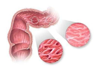
The anatomical structure of the colon is characterized by the existence of teniae, haustra and semilunar folds. The teniae are long, narrow bands of longitudinal muscle fibres, which are not equally spaced around the circumference of the colon, like in the small intestine, but make agglomerations. The colon walls are plicate: there are external bulges known as haustra, and the bulges to the intestinal lumen, referred to as semilunar folds.
The muscular coat (consisting of the longitudinal layer and the circular layer) is covered with the serous membrane from the outside, and the mucous membrane from the inside. Unlike the membrane of the small intestine, the mucous membrane does not have circular folds or intestinal villi, because its role is not to absorb food. On the other hand, in the mucous membrane of the large intestine there are more intestinal glands, which secrete big amounts of mucus – one of the components of stool.
The rectum
It is the final part of the colon. It consists of the pelvic part (the rectal membrane, located in the pelvis minor and the anus (the anal canal). These both parts together are about 17 cm long. The rectum differs slightly from the other parts of the large intestine. It does not have any teniae or semilunar folds, which are the characteristic features of the colon. Instead, in the mucous membrane we will find anal columns and transverse folds, which do not appear elsewhere. One of the important elements of the anatomical structure of the rectal wall is the internal anal sphincter, which, together with the external sphincter located below, fulfils a very important role in the process of defecation.
The large Intestine - Physiological Functions and Examination Methods
The basic function of the large intestine is to produce and secrete stool. Useless food residues (all the valuable elements are absorbed in the small intestine) are concentrated and processed by the bacteria of the large intestine.
If we suspect a colon disease, our diagnostic possibilities are much better than it is the case with the small intestine. Modern diagnostic methods, such as endoscopic tests, allow for a direct examination of possible pathological changes via a special instrument inserted per rectum into the intestinal lumen. The doctor who conducts the examination can see what our colon looks like and how it behaves. We differentiate two types of the examination, which differ in their place and scope. Rectosigmoidoscopy, which is an invasive instrumental examination, allows for the examination of the rectum and the final segment of the sigmoid colon. Colonoscopy enables the observation of the whole colon by means of optic instruments equipped with a light source. The first of these examinations is easier and can be done in ambulatory conditions. Colonoscopy, on the other hand, demands specialist skills and large experience and is harder to handle for the patient.
A traditional ambulatory examination is the radiography (x-ray), which consists of inserting some contrast media into the intestinal lumen and taking pictures. It is a valuable method complementing endoscopic examinations. It is also worth mentioning that there is a very simple examination per rectum, which in Poland is definitely preformed too seldom. In this test, the doctor inserts his finger into the anus and feels for abnormalities. Also laboratory blood tests and scatoscopy, including tests for unseen blood in the stool, are diagnostically important. At present, such tests can also be successfully preformed at home thanks to HemoActive rapid diagnostic tests produced by Biomerica. These tests are very easy to do by the patient alone and enable a frequent control of the large intestine. Using tests of this type allows for the detection of early warning signs of colorectal disease.
Colorectal Diseases
Unlike the small intestine, the large intestine is often affected by diseases, which can be very burdensome or life-endangering. The most common colorectal diseases include: hemorrhoids, colonic diverticula (especially of the sigmoid colon), appendicitis, intestinal obstruction, polyps or malignant tumors (cancer). Another common disease is IBS (Irritable Bowel Syndrome) which is characterized by a dysfunction of the large intestine.
The Colorectal Cancer – First of All: Early Diagnosis!
Statistics: The colorectal cancer incidence is growing systematically. It affects women and men alike.
According to the epidemiological data for 1996 there were:
- 5422 new cases in men (2732 – the colon, 2690 – the rectum)
- 3761 new cases in women (2033 – the rectum, 1728 – the colon).
The disease shows a constantly growing incidence, reaching the level of approximately 3,5% per year for men and about 2,5% for women.
The incidence is strictly connected with age and grows accordingly. It affects mainly men over 35 and women over 45 years old.
The curability of this disease is conditioned most of all by the early detection and quick treatment.
Factors contributing to the development of cancer:
Symptoms:
The disease development may go unnoticed. It is only in more advanced stages, when ailments connected with undernourishment and anemia appear, that the disease is discovered.
Clinical symptoms of colorectal cancer depend on its stage and location. Tumors located in lower parts may give the feeling of incomplete defecation, fecal incontinence and pain at defecation. Other symptoms may include:
Fortunately, the appearance of blood in the stool is often caused by hemorrhoids, while diarrhea or constipation may be caused by intestinal dysfunctions, such as the Irritable Bowel Syndrome.
Remember!

[maxbutton id=”15″]

[maxbutton name=”Kup online za 79,90 z?”]
W przypadku problemów z połączeniem naszych urządzeń z aplikacją Istel Health prosimy o kontakt: 800 70 30 11 lub mailowo: info@diagnosis.pl